| print
link to this post | email a friend Calcinosis Cutis & the Wonders of Laser Therapy!20 May 2022 By Laurie Edge-Hughes, BScPT, MAnimSt (Animal Physio), CAFCI, CCRT
I’ve had a couple of ‘let’s-try-it-and-it-worked’ cases in the past. I wanted to share the stories here, so that others might also benefit from what I learned.
Two past patients of mine presented with the diagnosis of Calcinosis Cutis. “What’s that”, you ask? Well, here are some links to bring you up to speed.
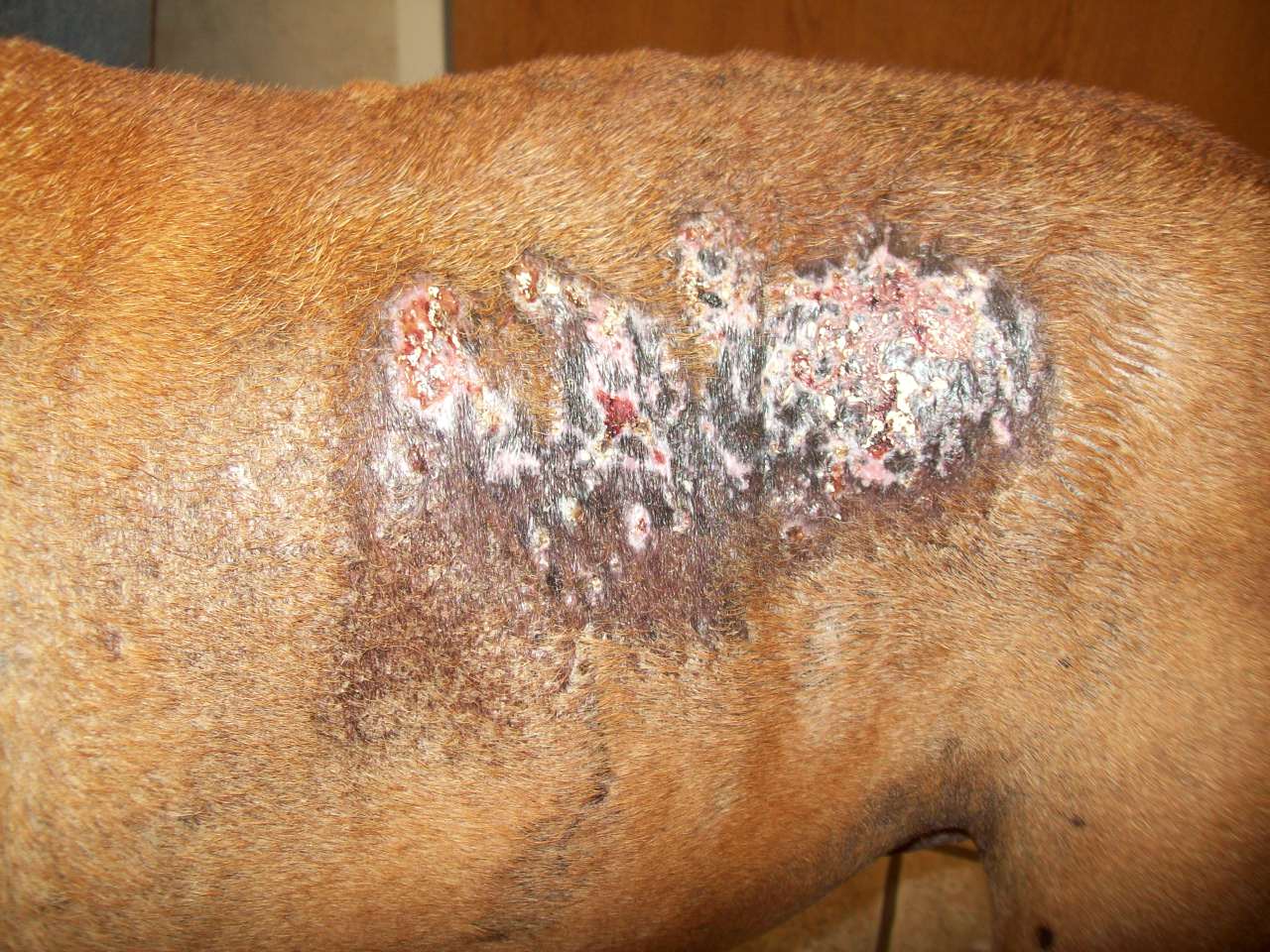
The description is as follows: Calcinosis cutis is the deposition of calcium in the skin, resulting from dystrophic or metastatic calcification. It is most commonly seen as a result of dystrophic calcification, which usually occurs more often in dogs than cats.
Dystrophic calcinosis cutis occurs in an area where there is damaged, inflamed, new growth or dead skin. Tissue damage may be from mechanical, chemical, infectious or other factors. Metastatic calcinosis cutis occurs in the setting of abnormal calcium and phosphate metabolism.
Widespread calcinosis cutis is referred to as calcinosis universalis, whereas more localized disease is called calcinosis circumscripta.
The most common underlying cause of calcinosis cutis is Cushing’s disease. Additionally, metastatic calcification can occur in dogs or cats with chronic renal failure.
In dogs, calcinosis cutis is often characteristic of Cushing's disease.
Other causes of cutaneous calcium deposition include:
Traditional veterinary treatment involves:
Case One of Calcinosis Cutis
The first case was a French Bulldog. He had come to the clinic following a cervical disc surgery. He had been put on prednisone for this issue and had subsequently developed several large patches of calcinosis lesions all over his body (back of his neck, over both shoulder regions and two patches on his back and flank area.
He was prescribed DMSO by his veterinarian to put on the lesions. His reason for coming to the clinic was because of his neck surgery… for which, he was doing quite well.
During his first appointment, I asked if we could do a trial using the laser on one patch of the calcinosis cutis. The owner was game to try. I chose to laser at a very low ‘wound healing’ dose (2.5J/cm2) using my Spectravet 810nm laser, 4 x 500mW multiprobe. [500mW, 2Hz, at 50% power, x 10 seconds]. I believe I lasered 5 or 6 points, which covered the entirety of the ‘test patch’ without any overlapping. (Sorry, memory is foggy and I’m not good about document how many spots I laser, however, I do remember wanting to be careful so as to not inadvertently flare up the condition… since this was just a test.)
The owner reported the following week that the test patch was remarkably better than any of the other patches. We subsequently treated again, but this time all patches at 5J/cm2. Again, improvement was seen in all areas lasered. The thickness of the patches was also beginning to normalize. Subsequent treatments were done at 10J/cm2. [Note: parameters as to Hz, power, and lack of overlap were the same as described above.]
Unfortunately, this dog died of unrelated cause before we were able full resolution. However, the trend was towards an overall improvement. The higher dose of laser seems to provide a favourable tissue response as did the lower dose. I kick myself for not taking pictures.
Case Two of Calcinosis Cutis
The second case was a wired haired dachshund. Her ‘human’ is a long-time client of mine and she had posted on Facebook asking if any of her dog-friends had any experience with calcium deposits in the foot pads or in the webbing between the foot pads. I posted one of the links above and asked if that was what she was referring to, and if so, that I had treated one dog where laser had improved them.
So, she booked her Daxie for an appointment and I lasered two areas on her paws (a thickened area in the main tarsal pad of a rear foot and another spot with several little ‘bumps’ in the webbing of a front foot.) As a side note, this little one had previously had surgical removal of the rear foot pad calcifications, but she was reluctant to do further surgery since the ones on the foot pad had come back (but weren’t currently painful). The owner reported that the dog would occasionally limp on the front foot. The lasering done for this little one consisted of the 4 x 500mW multiprobe head with the Spectravet laser, set at 2Hz, 50% power, for 40 seconds, thus delivering 10J/cm2. I did about 4 reps on each foot. Then I also lasered with the 904nm Superpulsed 4 diode multiprobe [Average Power output 50mW; Peak power output 25W] for 40 seconds on continuous wave, thus delivering 2J/cm2 with this device. Here again, I lasered each spot about 4 times. (I chose to use both laser heads because our single probe was out for servicing and the superpulsed head was able to get into the webspace of the front foot better than the 4 x 500mW 810nm head. Additionally, based on the readings I had done about calcinosis cutis, I advised the owner to do bloodwork at her vet to look for any potential underlying conditions.
The follow up appointment was 2-weeks later, and the owner was ecstatic. The lesions on both the front and the rear foot were dramatically improved. There had been no episodes of lameness either. (And bloodwork came back normal.) Treatment was the same on this day as it was the previous session. As well, her bloodwork had come back normal for all values.
A message a week after this treatment revealed that both sites were completely cleared, and that the owner would call if she noticed them re-appearing. _____
So, there you have it! I kick myself for not getting pictures of ANY of these lesions! However, I hope the story will lodge in your brain to pop up when needed should you ever have a dog with Calcinosis Cutis!
|
|