| print
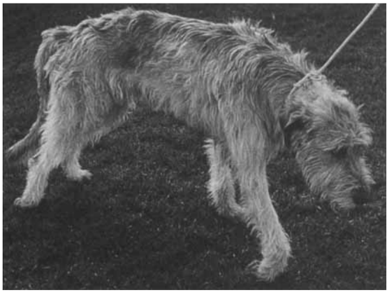
link to this post | email a friend Fibrocartilaginous Embolism – A REWARDING CONDITION FOR REHAB18 Jun 2020 By Margaret Kraeling, PT, CCRT Fibrocartilaginous embolic myelopathy (FCEM) is a common and very rewarding condition to treat in the canine rehabilitation clinic.
Fibrocartilaginous embolic myelopathy is a vascular disease of the spinal cord caused by embolization of spinal vasculature with fibrocartilaginous material histologically and histochemically identical to the nucleus pulposus of the intervertebral disk, resulting in ischemic necrosis of dependent regions of spinal cord. Various theories have been hypothesized to explain how the fibrocartilaginous material can enter the spinal vasculature. FCEM is considered as the most common cause of ischemic myelopathy in dogs. There is no clinical test currently available to absolutely confirm a diagnosis of a fibrocartilaginous embolism (FCE). Initially the aim is to differentiate between FCEM and other conditions such as intervertebral disc disease, a fracture, or a spinal tumor -- all of which can cause considerable pain. If the dog is not in pain, that is one factor that can be indicative of an FCE. The best imaging technique for a fibrocartilaginous embolism is the MRI, because it can distinguish between an obstruction (i.e. an embolism) and a compression or swelling of the spinal cord. Neurologic signs are peracute in onset and the severity and distribution of the signs are directly in relation to the site and the extent of spinal cord infarction. The neurologic signs usually stabilize in the initial 24 hours and then may remain static or improve depending on the severity and the extent of the ischemia and speed in which rehabilitation is instituted. FCEM is considered as the most common cause of ischemic myelopathy in dogs. The general thought is that the severity of neurological signs at the time of initial examination and MRI (if used), are associated with the outcome. The variable in this outcome however was seen when physiotherapy including hydrotherapy was instituted immediately after the diagnostic work-up. These cases saw a major improvement in recovery rate and overall function. Another study reported no additional benefit when comparing dogs treated with physiotherapy alone versus those treated with physiotherapy and corticosteroids. FCE development has been reported predominantly at the vertebral levels C6-T2 and L4-S3. It is noted that there is a positive correlation between a poor prognosis and the involvement of intumescences, symmetrical clinical signs and decreased deep pain sensation. However, physiotherapy and/or hydrotherapy instituted immediately after the diagnosis seems to have a major influence on the recovery. One study indicated that early precise diagnosis as well as early onset of treatment are important to obtain a favorable prognosis. This study noted treatment commenced in less than 15 days. In dogs with FCEM, time intervals between onset of neurological signs and recovery of voluntary motor activity, unassisted ambulation, and maximal recovery have been reported as varying between 6 days and 3.75 months. One study reported that 88% of the dogs were small to medium sized which is not the usual demographic that is reported. Most commonly this condition has been reported in large and giant breed non-chondrodystrophic dogs. However, it has also been described frequently in small breed dogs (particularly miniature schnauzers). The male to female ratio in dogs ranges from 1:1 to 2.5:1 in different studies. The age at diagnosis is an average of 4-6 years old. Onset of neurological dysfunction during physical activity, such as running, jumping, or playing, is common but occasionally no triggering event is noted. Marked lateralization of neurological dysfunction has been reported as 53–87% in various studies on FCEM. Treatment Treatment will vary with each case and will depend on a thorough evaluation when the dog presents for rehabilitation. We have seen dogs as early as 24-hours post onset. Treatment can range from initial passive range of motion, bed positioning and instruction to manage activities of daily living (ADL) for the owners such as lifting aides like the ‘Help Em Up’ harness, addressing bowel and bladder routines, and prevention of pressure sores. A wide range of modalities can be used for these dogs from laser, pulsed magnetic field, microcurrent, acupuncture and electrical muscle stimulation, all depending on the findings at evaluation. There will also be very specific therapeutic exercises which will be tailored to each case and will be ever changing throughout the progression of recovery. Both pool and underwater treadmill can be extremely useful at varying times during the course of recovery as well.
Because these dogs usually have an excellent prognosis the owners are eager to become involved in their home exercise programs and to help their pet reach maximum functional recovery. A fun and rewarding case for all involved. References 1.De Riso L & Platt SM. Fibrocartilaginous Embolic Myelopathy in Small Animals. Vet Clin Small Anim 40 (2010) 859–869.2.De Riso L, Adams V, Dennis R et al. Association of clinical and magnetic resonance imaging findings with outcome in dogs suspected to have ischemic myelopathy: 50 cases (2000–2006). J Am Vet Med Assoc. July 1, 2008, Vol. 233, No. 1, Pages 129-135.3.Nakamoto Y, Ozawa T, Katakabe K et al. Fibrocartilaginous Embolism of the Spinal Cord Diagnosed by Characteristic Clinical Findings and Magnetic Resonance Imaging in 26 Dogs Journal of Veterinary Medical Science 2009.4.Nakamoto Y, Ozawa T, Katakbe K et al. Usefulness of an early diagnosis for the favorable prognosis of fibrocartilaginous embolism diagnosed by magnetic resonance imaging in 10 Small- to Middle-Sized Dogs. Veterinary Research Communications May 20, 200.5.De Risio L. A review of fibrocartilaginous embolic myelopathy and different types of peracute non-compressive intervertebral disk extrusions in dogs and cats. Front. Vet. Sci., 18 August 2015.6.Gandini G, Cizinauskas S, Lang J, et al. Fibrocartilaginous embolism in 75 dogs: clinical findings and factors influencing the recovery rate. J Small Anim Pract. 2003 Feb;44(2):76-80.
|
|