| print
link to this post | email a friend Clyde – A most fascinating case!!!28 Oct 2019
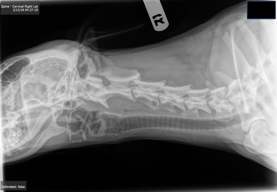
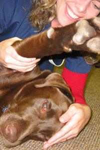
By Margaret Kraeling, PT, CCRT Clyde, a male border collie, was just past one year old when he was first seen in our clinic on February 7, 2018. He presented with unilateral facial atrophy on the left side. He was also seen at the vet for fever and lethargy. He had a previous episode of lethargy in mid-December. At that time he did not want to bend down to eat. Prior to this episode he was a normal active puppy. He was at that time being treated with antibiotics and meloxicam and was awaiting results from recent testing for cytokines. The owner reported that Clyde played very rough with the other young dog in the household – usually not coming out on the winning side! The other dog would typically grab him by the collar and pull him down to the ground. There was also a report of a collision with the other dog at the dog park about 4 weeks prior to our appointment. When I first saw Clyde he was extremely lethargic – most unlike I would have expected from a young border collie. When I tried to interest him in a ball he crouched down to pick it up cautiously and it seemed that neck movement was uncomfortable and restricted. The cervical spine was examined cautiously as no x-rays had been taken at this point. C1 was palpated to be in RSB / LR; C7 was tender and there was a 50% decrease in RSB. Restrictions in motion (via craniosacral osteopathic evaluation) at the cranial sutures was noted at temporal / parietal / occiput and sphenoid / occiput as compared to the right side. There was atrophy of temporalis and masseter muscles on the left side however he did have control of eye movements as well as opening and closing. The temporalis and masseter responded to E-Stim. At that initial appointment I treated him with gentle manual therapy, and laser (both to the cervical spine, and acupuncture points ST6, ST4, GB1, TW17) as well as E-Stim to the facial muscles. In addition I used some craniosacral techniques to restore symmetrical motion on both sides of the cranium. Clyde was followed up on Feb. 12 at which time he appeared somewhat more energetic. He was no longer on antibiotics or metacam and his fever had resolved. The cytokine testing was reported as normal. He did appear to have some trouble swallowing food so the dishes were elevated for him. Similar findings in cervical joint positioning and restrictions were evident. At this appointment, as was common in other appointments, Clyde fell asleep during the manual therapy portion. On February 13 the owner called to say there was good response from treatment and Clyde was able to get comfortable at night to sleep rather than being very restless which had been the case. She did, however, notice that his left pupil did not appear to be reacting to light as the right side was. She was advised to return to see her vet who did x-rays and organized a consult with ophthalmologist. At that time the reflexes were all normal but he suggested there may have been some pressure on the oculomotor nerve although he did not offer a cause for that. He was prescribed steroids and gabapentin prior to re-assessment. By February 15 the pupil was reacting normally and equal to the right. He was eating well with no further episodes of difficulty swallowing. February 20 Clyde attended for another follow-up in our clinic at which time his owner reported that he wasn’t blinking as much in his left eye and therefore had now developed a corneal ulcer. Drops were prescribed by his veterinarian. C2 was palpated to be in LR. There appeared to be less atrophy in the facial muscles and he was able to use masseter and temporalis when taking a cookie from the left side of the mouth. Treatment today was modalities only as we were concerned there may be some increased space at C1/C2 and we encouraged the owner to have the x-rays sent out to a veterinary radiology specialist. Clyde’s next follow up was March 12. The owner reported that they have had a difficult time trying to find eye drops that did not cause an adverse reaction for Clyde. He was also continuing to wean off the steroids. The x-rays have been reported as normal. The left eye still had an opaque film on it however the eyelid could now close and the blink reflex was improving. Treatment continued with E-Stim to facial muscles and improved voluntary control was noted following. Palpation of C0/C1/C2 showed good position and there was no muscle spasm along the neck on either side. Cranial bones were also moving symmetrically on both sides as per his craniosacral evaluation. Two more follow ups were done on April 21 and May26 at which time continued improvement was noted. By May 26 the muscle bulk of masseters and temporalis were almost equal to the right and there was just slightly less elevation of the eyelid on the left. There were still some minor restrictions in the cervical spine which were addressed at those appointments. There was one reported relapse in June again after Clyde was playing with his housemate. He was seen at the vet for fever and treated with antibiotics. By mid-July Clyde appeared completely recovered. The corneal and nasal reflexes were equal bilaterally. Cervical spine motion was equal bilaterally and facial muscles were symmetrical. Clyde’s owner is supervising all play with the other dog in the house and rough play is not permitted. Clyde no longer wears a collar in the house and when out for walks he uses a harness. Below is a copy of the x-ray that we were concerned about. It was never sent to a veterinary radiology specialist however was reported as normal by two or three regular vets that had seen Clyde during this period.
We never had an actual diagnosis on this dog as is common in our field. The first impression from the vet was MMM (masticatory muscle myositis) however both the owner (previously a vet tech) and myself questioned that as Clyde’s symptoms were unilateral, the muscles were not swollen and painful and he could open his mouth. The vets were also testing for other immune mediated diseases – all the tests that were done revealed nothing to indicate an autoimmune response. As the dog gradually recovered there was no further attempt to offer a diagnosis. As we looked at the x-ray and considered the anastomoses between the lower cranial nerves (particularly facial, trigeminal and hypoglossal) and the upper cervical nerves we still considered that trauma was the likely cause.
|
|